CHOC is leading a new international study to determine if cardiac magnetic resonance imaging (MRI) can help surgeons choose between the more desirable of two surgical options for patients born with congenital heart diseases involving underdeveloped left ventricles.
This relatively rare group of conditions presents two main surgical options: biventricular, or two-ventricle, repair, the more desirable choice; or single-ventricle repair, which is more complex and eventually leads to complications and heart transplantation.
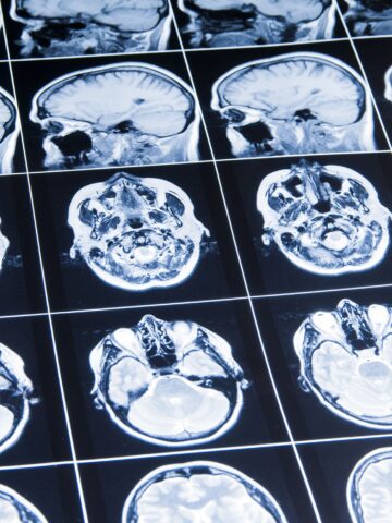
Picking the best option for a patient is crucial to a patient’s long-term outcome but selecting which option to pursue can be challenging. The choice often relies on cardiovascular imaging techniques such as echocardiography and, more recently, cardiac MRI.
CHOC, UCLA Health affiliation
The new study, called BELIEVE-CMR (Borderline Left Ventricle Evaluation with Cardiac Magnetic Resonance), is an example of how CHOC and UCLA Health are transforming the way cardiac care is being delivered in Southern California. In 2023, the two pediatric healthcare systems launched a joint pediatric heart program.
The patients in the new study have “borderline” left ventricles (BLV), meaning the size of their left ventricles does not clearly lead to a clear consensus on which surgery is the most ideal to perform.
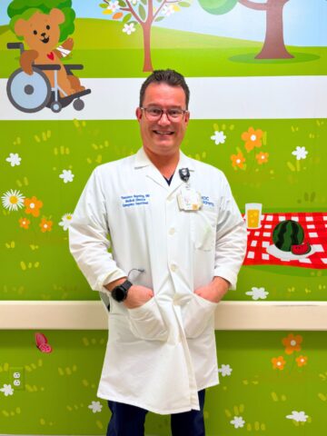
Dr. Glen Van Arsdell, chief of the CHOC/UCLA Health pediatric heart program and chief of congenital cardiovascular surgery at UCLA Health, contributed pioneering research involving the benefits of using cardiac MRIs to assess BLV patients.
In his smaller, single-hospital studies, state-of-the-art MRI technology has given cardiac specialists the best possible views of the left ventricle, which is expected to pump blood to the whole body.
“Without the MRIs,” Dr. Van Arsdell says, “it’s much more of a guess.”
15-site study
The BELIEVE-CMR study hopes to involve 15 pediatric hospitals across the U.S. and around the world, with an expected patient enrollment of 200. The research could help provide a definitive answer as to whether cardiac MRIs should become the preferred tool to guide the decision of which surgical option to pursue for this subset of congenital heart disease patients.
“The MRI might be the decision-maker,” agrees pediatric cardiologist Dr. Pierangelo Renella, the primary investigator of the BELIEVE-CMR study and director of the Cardiac MRI Program at CHOC.
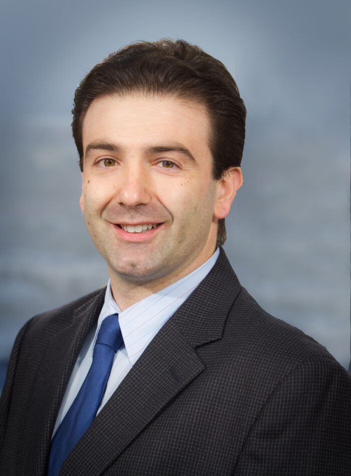
“Cardiac MRI is currently the only imaging method that can provide the level of detail needed to determine the size and function of the left ventricle, as well as the amount of blood it can carry,” Dr. Renella explains. “Our study will look at how MRI data might predict patient outcomes, which may allow us to choose which patients may be ‘rescued’ from the fate of having to undergo a single-ventricle procedure.”
Several patients at CHOC and at UCLA, from newborns to 5-year-olds, are already enrolled in the study, which is funded by the CHOC Heart Institute and the CHOC Chief Scientific Officer small grants program.
Because BLV cases aren’t that common the study is expected to last a few years, Dr. Van Arsdell says.
Expertise required
Traditional methods of getting inside views of the heart include echocardiograms, CT scans, chest X-rays, and cardiac catheterization. Some of these techniques involve exposure to radiation and/or do not provide the needed details to move forward with a given surgical option.
“What MRI does for us is it gives us functional data. which really helps us see not only what a heart looks like, but what it can do,” Dr. Van Arsdell explains. “Echocardiograms don’t tell you quite as much about what the heart can do, so MRIs help us make better decisions.
“Our job is to determine if the ventricle is too small and needs to be a one-ventricle repair, or if it’s borderline, and if so, we then can grow it and it can become a two-ventricle repair,” he adds. “The MRI really helps us with the borderline cases. We can then adopt a strategy to disproportionally grow the ventricles and the valves so we can turn them into a two-ventricle repairs.”
Some challenges
Although cardiac MRIs could be deemed the most important imaging method to assess BLV patients, and potentially aided by artificial intelligence (AI), the technology is relatively more costly than, for example, echocardiograms, which use sound waves to create pictures of the heart, Dr. Renella notes.
Cardiac MRIs may also require general anesthesia in younger patients and the expertise needed to interpret them is less widely available, Dr. Renella says.
But the CHOC-UCLA Health affiliation is poised to become a leader in using cardiac MRIs to help surgeons decide on how to repair defects in BLV patients with an optimal way to measure ventricle size and function, amount of blood flow, blood vessel size, heart valve anatomy, and scar tissue in the heart muscle, Dr. Renella says.
“Cardiac MRIs can collect a wealth of information about the hearts of our most complicated cardiac patients,” Dr. Renella says. “It is now our job to decode which pieces of that information help those patients do better after surgery.”
CHOC and UCLA Health together have been ranked among the top children’s hospitals in the nation for Cardiology & Heart Surgery by U.S. News & World Report.