For pediatric neurosurgeon, Dr. Suresh Magge and his colleagues in the CHOC Neuroscience Institute, Christmas this year arrived in late June.
That’s when a 3D camera system was installed in the neurosurgery clinic that will significantly advance CHOC’s mission to treat craniosynostosis, says Dr. Magge, PSF neurosurgery division chief for CHOC and co-medical director of the Neuroscience Institute.
Craniosynostosis, which affects 1 in every 2,000 infants, causes an infant’s skull to fuse early, creating an irregular skull shape, and can lead to increased pressure on the brain as a child matures. This can lead to headaches, vision problems, and cognitive issues.
The 3D motion-capture camera can, in seconds, capture a comprehensive array of images that will allow CHOC neurosurgeons to better analyze and measure in detail a child’s head. This, in turn, will allow them to enhance research in craniosynostosis and design the best possible treatments.
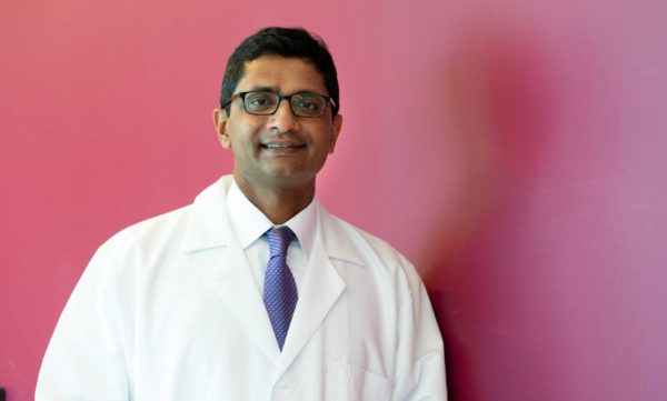
“This really makes a difference,” says Dr. Magge, noting that traditional 2D photos and measurements “only go so far.”
“This new camera allows us to get data quickly and safely,” Dr. Magge says.
CHOC recently had its first craniosynostosis patient imaged by the 3D camera.
A push to greatness
The new camera is a critical step in Dr. Magge’s push to advance the path of the Neuroscience Institute in becoming a world-class destination for neurological care.
Dr. Magge was recruited to CHOC last October after an 11-year tenure at Children’s National Hospital in Washington, D.C., where he started the medical center’s neurosurgery fellowship training program and was the director of medical student education in pediatric neurosurgery.
During his time at Children’s National, Dr. Magge started the region’s first minimally invasive craniosynostosis program. He aims to launch a similar program at CHOC as well.
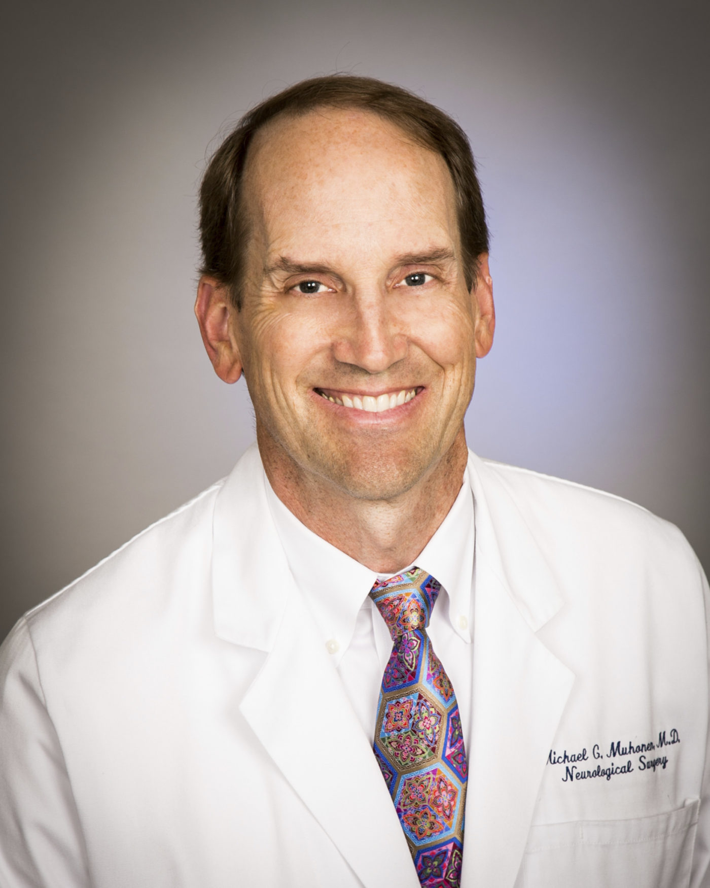
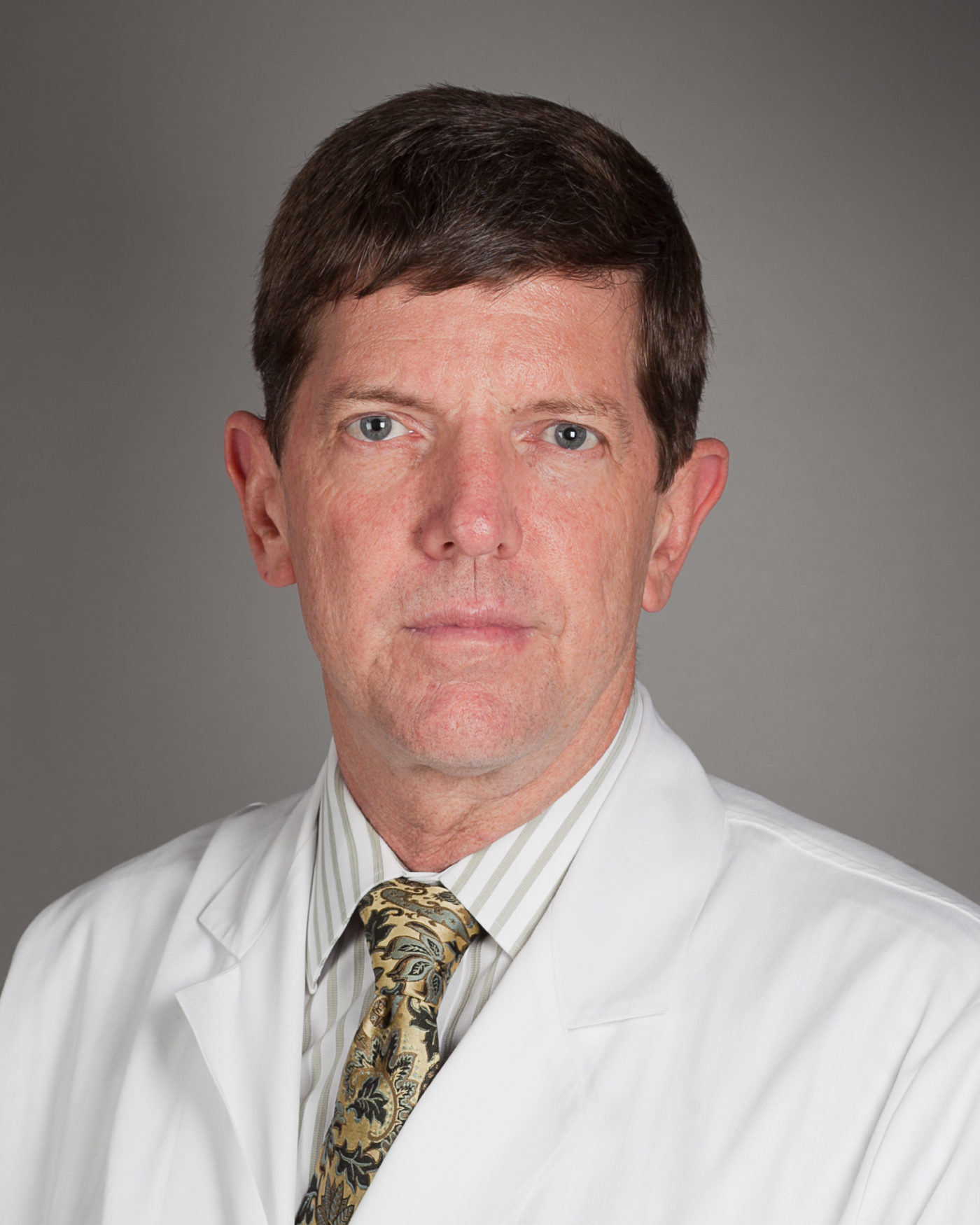
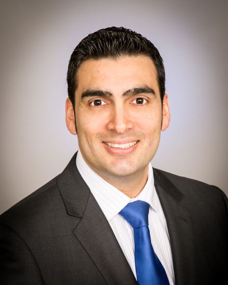
The neurosurgery division also includes Dr. Michael Muhonen, Dr. William Loudon, and Dr. Joffre Olaya.
For the last several decades, the go-to surgery to treat craniosynostosis has been an open surgical correction called calvarial reconstruction. For this surgery, doctors must wait until the child is 6-12 months of age and perform a large surgery that involves opening the scalp, taking apart the entire skull, then putting it back together.
“It’s a good surgery, and most kids do well, but we have newer techniques that are less invasive,” Dr. Magge says. The open calvarial reconstruction surgery generally takes 4-6 hours and requires a hospital stay of 3-5 days, as well as a blood transfusion during surgery.
Unlocking the skull
The minimally invasive procedure Dr. Magge learned during his fellowship at Boston Children’s Hospital involves using an endoscope with a camera attached to its tip.
After making one or two small incisions, Dr. Magge goes under the scalp and then under the skull, using the endoscope to separate the skull from the underlying tissue. He then cuts out a piece of bone — 1 to 2 centimeters in width – to “unlock” the skull.
This surgery only takes about an hour, and most children don’t need a blood transfusion and can go home the next day. After surgery, they wear a molding helmet that helps to reshape the skull. This minimally invasive surgery is generally done by 3-4 months of age (earlier than the open surgery).
“The data shows this surgery works very well,” says Dr. Magge, who has given many presentations and written multiple papers about this procedure.
The aesthetic results have been shown to be excellent in many papers, Dr. Magge says. What still needs to be verified by research are the long-term cognitive outcomes of patients after either the open or minimally invasive surgery.
To that end, Dr. Magge launched a study about two years ago involving patients from multiple hospitals that looks at children five years after surgery. Dr. Magge plans to enroll patients from CHOC in the study.
He estimates the study will be completed in about two years.
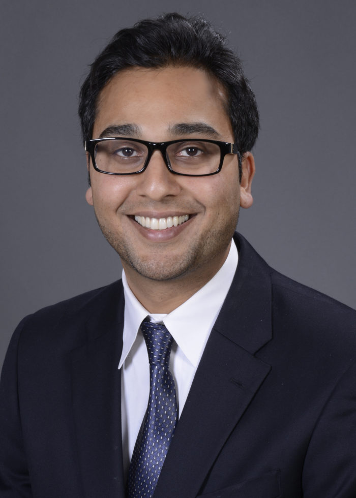
Working with plastic surgeon Dr. Raj Vyas, Dr. Magge says CHOC offers comprehensive cranio-facial services.
“To be comprehensive,” he says, “you have to offer traditional surgery as well as minimally invasive surgery.”
Part of the funding for the new 3D camera came from a CSO grant established by CHOC Vice President for Research and Chief Scientific Officer Dr. Terence Sanger, a physician, engineer, and computational neuroscientist who also is vice chair of research for pediatrics at the UCI School of Medicine.

The 3D camera arrives as significant infrastructure changes are underway at the Neuroscience Institute: CHOC recently opened its new state-of-the-art outpatient center, establishing a clinical hub for caregivers to serve patients and families in a centralized location. Additionally, plans are underway to expand the hospital’s inpatient neuroscience unit.
“CHOC has been very supportive of the Neuroscience Institute,” Dr. Magge says. “I’m very excited.”

CHOC Hospital was named one of the nation’s best children’s hospitals by U.S. News & World Report in its 2025-26 Best Children’s Hospitals rankings and ranked in the neurology/neurosurgery specialty.

