For several weeks, pediatric neurosurgeon Dr. Aaron Yengo-Kahn kept the printed 3D model of a toddler’s spine on his desk at Rady Children’s Hospital.
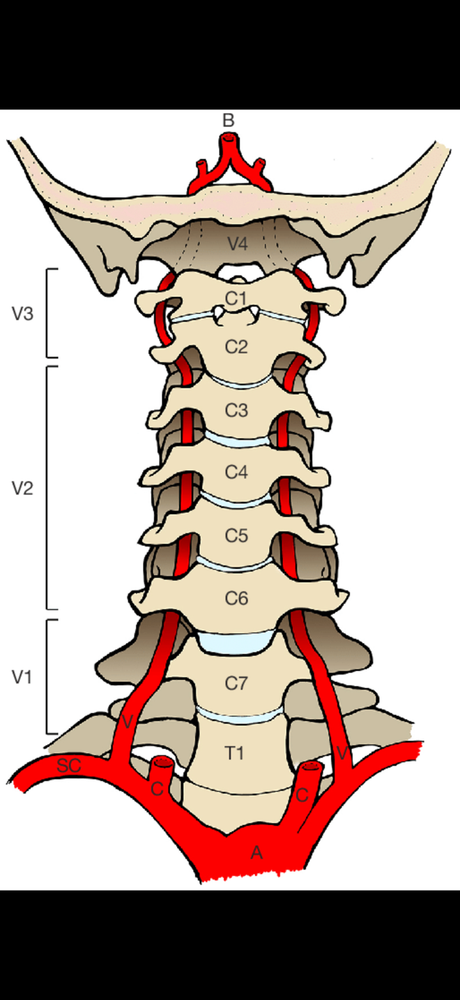
He took it home to memorize every detailed contour of the 7-inch model, which also depicted in bright red the blood vessels and aortic arch.
Come surgery day, Dr. Yengo-Kahn placed the model in a sterile plastic bag for reference as he took a scalpel to the neck and back of his 2-year-old patient to delicately cut, reshape and realign bones and fuse them with rods and screws to protect the child’s spinal cord.
Born with a condition that caused severe craniocervical instability, the boy’s underdeveloped bones and ligaments continuously put his spinal cord function at risk, requiring the use of a neck brace almost since birth.
The internal hardware – some of it temporary – was designed to give him more stability to protect his spinal cord from further injury and allow for recovery.
The best planning tool possible
Although 3D-printed models are not new to pediatric healthcare, Rady Children’s Hospital Orange County now has four of its own printers in the new maker space in the Research Institute on the ninth floor of the Southwest Tower.
Until this March, surgeons had to outsource the work.
Research scientist Sumiko Abe, supervisor of advanced imaging intelligence at Rady Children’s Hospital Orange County, specializes in advanced medical imaging and computational neuroimaging. She converts patients’ CT and MRI data into detailed, patient-specific 3D anatomical models, carefully segmenting structures such as bone and blood vessels before preparing them for printing.
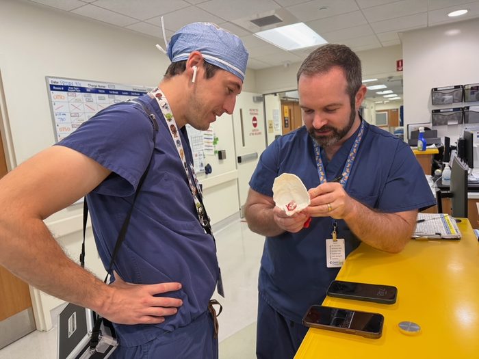
Since June, Sumiko has made more than a dozen models for surgeons like Dr. Yengo-Kahn, who says they help him get the best possible realistic views of a patient’s anatomy prior to surgery.
That, in turn, allows him to come up with the most effective surgical procedures – especially for those with complex anatomical conditions like the 2-year-old spinal patient who was born with congenital stenosis of the spinal canal and odontoid hypoplasia contributing to instability.
Pediatric neurosurgeon Dr. Joffre Olaya, division chief of neurosurgery, performed surgery on the boy when he was a newborn to relieve the spinal cord compression but as he grew, head and neck instability remained a concern, so doctor equipped him with a stabilizing collar to temporize him until he was big enough to undergo surgery with spinal instrumentation.
Sumiko observed part of the recent surgery.
“Dr. Yengo-Kahn has very challenging cases, and I love being able to create these tools to help him plan the best surgeries,” she says.
Looking inside the body
Dr. Yengo-Kahn explains the value of printed 3D models.
“At the crux of complex spinal surgeries is a very physical, biomechanical problem that’s really hard to visualize, even with 3D computerized renderings,” he says. “And it’s nearly impossible to make sense of congenital anatomical anomalies using just 2D images.”
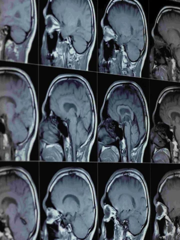
Getting a look inside bodies starts with computed tomography (CT) scans, which combine specialized X-ray equipment with computer processing to create detailed cross-sectional pictures. But there are limitations.
“CT scans can show us the bone anatomy and help us select screw sizes and get a basic understanding of the surgery, but you need to understand a lot more than that – such as where you need to move the spine, for example.
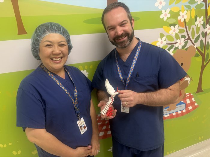
“With these 3D printed models, we can show up to the OR and not just have a plan we made on paper, but a full 3D anatomy in our heads of the specific needs of each patient. This is the best surgical navigation tool you can have, and the models Sumiko creates are incredibly accurate.
“She is super talented with this stuff, and since this is her job, we’re not using up surgeons’ time to create them.”
A gift
Dr. Yengo-Kahn’s six-hour surgical procedure in mid-September went very well. Pediatric orthopedic spine surgeon Dr. Taylor Jackson,who cares for babies, kids, and teens with complex musculoskeletal conditions, assisted him, highlighting the collaborative relationship between neurosurgery and orthopedic surgery in treating complex spinal pathology at Rady Children’s Hospital Orange County.
The boy will wear a stabilizing collar for added support for a few months.
In about four to five months, Dr. Yengo-Kahn will remove the temporary rods and screws, leaving just the permanently fused hardware from the base of the skull to the second vertebrae in his neck to keep the boy’s upper spine stabilized.
Sumiko, who joined the hospital formerly known as CHOC in 2020, expects to keep busy making the models for a growing number of service lines.
“We’re at the forefront of doing this among pediatric hospitals in California,” she says.
After the surgery, Dr. Yengo-Kahn gave the boy’s parents the 3D model.
Looking ahead
Dr. Yengo-Kahn says printed 3D models, which typically are made of various plastics, liquid resins, metals, and composite materials, are amazing for planning and inter-operative work.
However, they are expensive to make.
He’s hopeful a donor will step forward to allow Rady Children’s Hospital Orange County to continue using them as often as possible and allow for eventual upgrades to print progressively more complex pathology including the brain and spinal cord for congenital malformations and tumors.
“This effort falls in line with our vision of becoming the most comprehensive spine center in Southern California,” Dr. Yengo-Kahn says. “Surgeons have much smoother cases when these models are used, and parent and patient satisfaction is so much higher.”
For years, internationally recognized pediatric orthopedic surgeon Dr. Afshin Aminian, medical director of the Orthopedic Institute at Rady Children’s Hospital Orange County, has incorporated 3D printed models in his work.
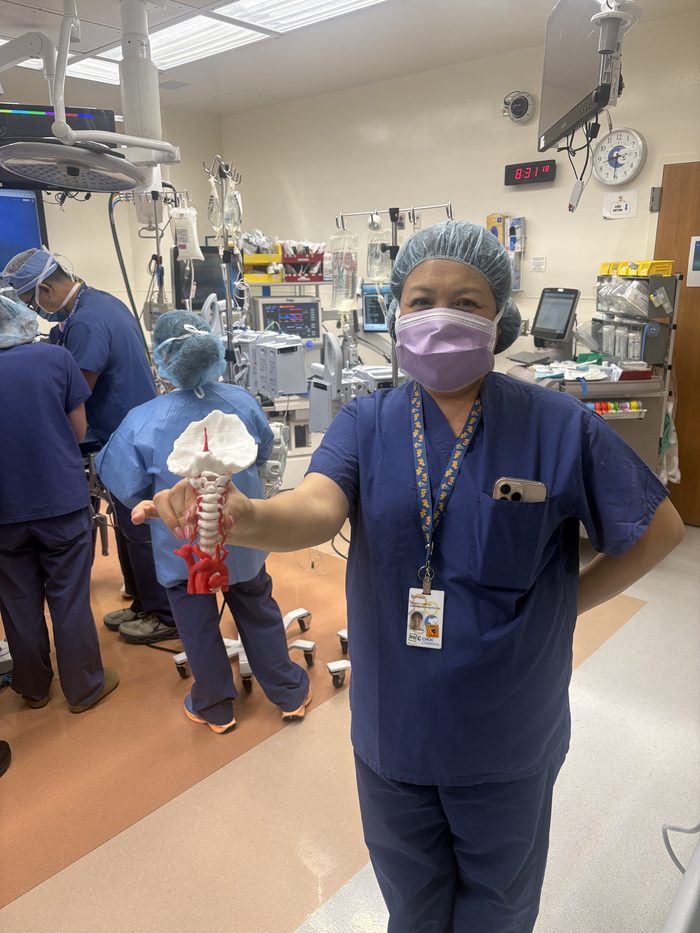
Together, Dr. Aminian and Dr. Yengo-Kahn have tackled some of the most complex cases of kids with spinal deformities — not only correcting structural alignment but also improving neurological functioning. Having the four 3D printers on site helps streamline their partnership, which is uncommon for a pediatric hospital.
Looking ahead, Sumiko sees 3D printing as one part of a broader patient-specific modeling program at Rady Children’s Health. The same advanced imaging and modeling techniques used to create physical models can also support digital and emerging visualization technologies, expanding their potential use across surgical specialties, emergency medicine, education, research and other areas of pediatric care, she says.
“The real foundation is the ability to transform a child’s medical images into an accurate, patient-specific 3D model,” Sumiko says. “Today the model may be something a surgeon can hold in their hands. In the future, the same model could also be used in advanced digital or virtual environments. We’re building something that can grow well beyond 3D printing. The long-term vision is to create a patient-specific modeling platform that can support many specialties across pediatric care.”
Refer to a CHOC Pediatric Neurosurgery Specialist
Referrals can be placed online through the eCeptionist Referral Portal.